
Radguide
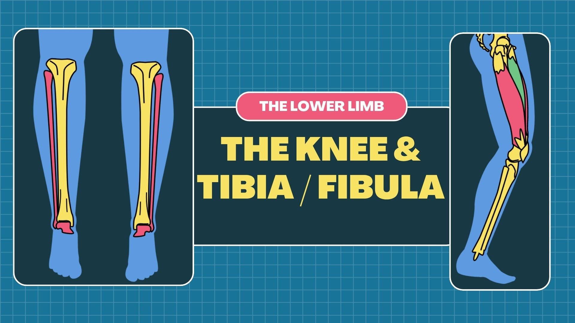
Tibia - Fibula & Knee : Osteology & Radiographic Technique
Tibia - Fibula & Knee : Osteology & Radiographic Technique
Couldn't load pickup availability
Master Tibia-Fibula & Knee Imaging: Osteology & Radiographic Techniques
Unlock the structural complexities of the tibia, fibula, and knee joint with RadGuide’s detailed, downloadable presentation. Ideal for radiography students, healthcare professionals, and anyone looking to enhance their skills in lower limb imaging, this resource offers an in-depth look at the anatomy of the tibia, fibula, and knee, along with essential radiographic techniques to ensure accurate, high-quality diagnostic images.
📘 In this presentation, you’ll explore:
Tibia-Fibula & Knee Anatomy: Osteology & Key Landmarks
🦴 Tibia-Fibula Osteology Overview
-
Comprehensive breakdown of the tibia (shinbone) and fibula, highlighting their structure and functions in weight-bearing and mobility
-
Key anatomical features of the tibia: the tibial plateau, medial malleolus, and tibial tuberosity
-
Understanding the fibula's role in support, with focus on the lateral malleolus and distal fibular articulation with the tibia
-
Visual aids that clearly depict the proximal tibiofibular joint, distal tibiofibular joint, and their articulations with the knee and ankle
📏 Knee Joint Anatomy
-
Detailed anatomy of the knee joint, including the femur, tibia, fibula, and patella
-
Study of key knee structures such as the femoral condyles, tibial plateau, menisci, and ligamentous structures (ACL, PCL, MCL, LCL)
-
Visual aids to understand the relationship between the knee joint and surrounding muscles, tendons, and ligaments
Radiographic Techniques for the Tibia, Fibula & Knee
📸 Standard Projections for Tibia-Fibula & Knee Imaging
-
Step-by-step guidance on common radiographic views for the tibia and fibula: AP, lateral, and oblique projections for both the tibia and fibula
-
Detailed instructions on knee projections, including AP, lateral, sunrise (merchant view), and bilateral weight-bearing views
-
Special techniques for knee arthrograms, patellar fractures, and joint space evaluations
🎯 Optimizing Radiographic Technique
-
Tips for adjusting exposure factors (kVp, mAs) to achieve high-quality tibia, fibula, and knee radiographs with minimal distortion
-
Best practices for centering points and beam angles to capture both tibia-fibula alignment and knee joint detail
-
Key positioning techniques to ensure accurate visualization of bone structures, joint spaces, and ligamentous structures in radiographs
💡 Positioning for Clinical Indications
-
Techniques for imaging common knee injuries, including fractures of the tibia and fibula, dislocations, and ligament injuries
-
Radiographic strategies for assessing joint spaces, arthritis, and degenerative joint disease in the knee
-
Methods for imaging meniscal tears, osteochondral defects, and other soft tissue injuries related to the knee joint
Educational Value and Application
Designed for:
-
Radiography students learning lower limb anatomy and radiographic techniques
-
Healthcare educators teaching musculoskeletal anatomy and imaging in clinical settings
-
Radiography professionals seeking to refine techniques for imaging tibia, fibula, and knee injuries
Format: Digital Presentation (PowerPoint/PDF)
Delivery: Sent via email upon purchase
Level: Beginner to intermediate
Created by: Registered radiographer with clinical teaching experience
Share
