
Radguide
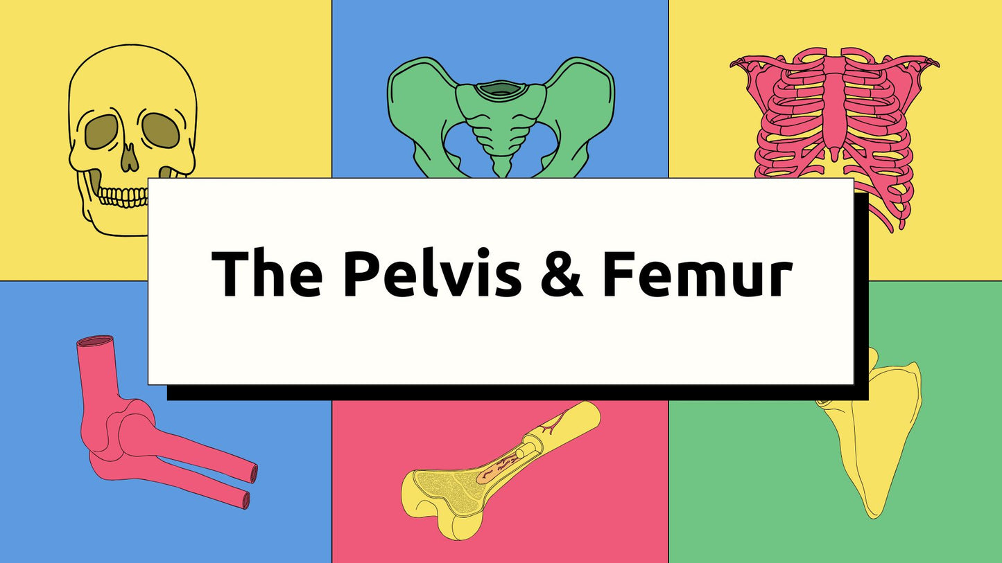
The Pelvis & Femur : Osteology & Radiographic Technique
The Pelvis & Femur : Osteology & Radiographic Technique
Couldn't load pickup availability
Build a Strong Foundation in Pelvic Osteology, Anatomy & Radiographic Technique
Explore the structural complexity of the pelvis with RadGuide’s comprehensive digital presentation. Perfect for anatomy students, radiography trainees, and healthcare professionals, this resource offers a clear and concise introduction to the bones and landmarks of the pelvic region, essential for understanding movement, clinical assessment, and imaging. In addition, gain a strong foundation in pelvic radiographic techniques to ensure accurate imaging and diagnosis.
📘 In this presentation, you’ll explore:
Pelvic Osteology Overview
🦴 Detailed Breakdown of the Pelvic Bones
-
Thorough examination of the bones that make up the pelvis: ilium, ischium, pubis, sacrum, and coccyx
-
Understanding how these components fuse to form the hip bones, supporting the spine and lower limbs
-
Key anatomical features such as the iliac crests, anterior superior iliac spine (ASIS), and acetabulum
📏 Key Anatomical Landmarks
-
Identify important features including the pubic symphysis, sacroiliac joints, and pelvic inlet/outlet
-
Clinical significance of anatomical landmarks such as sacroiliac joints for assessing conditions like arthritis and inflammation
🔍 Pelvic Structure and Function
-
Insight into the role of the pelvis in weight transfer, locomotion, and organ protection
-
The functional demands of the pelvis, including its role in childbirth
-
Divisions of the true and false pelvis, and the anatomical relationships that allow for proper movement and posture
Anatomical Relationships and Joints
💡 Pelvic Joints & Articulations
-
Explore how the pelvic bones articulate with the spine and femurs, forming essential joints like the sacroiliac (SI) joints and hip joints
-
Understand how these joints contribute to posture, gait, and functional movement
-
Study the pelvic inlet and outlet, their relationship to the birth canal, and implications for obstetric imaging
Radiographic Technique for Pelvic Imaging
📸 Pelvic Radiographic Projections
-
Step-by-step guidance for key pelvic imaging views: AP (Anteroposterior), lateral, Judet views, and oblique views for specialized imaging
-
Proper positioning techniques to ensure accurate alignment and visualization of pelvic bones and joints
-
Specific instructions for hip joint imaging and fracture detection
🎯 Optimizing Radiographic Technique
-
Best practices for adjusting exposure factors (kVp, mAs) to obtain optimal image quality for bone detail and soft tissue visibility
-
Key centering points and beam angles for accurate radiographs of the pelvis and hip joints
-
Ensuring proper patient positioning to avoid distortion and superimposition of anatomical structures
💡 Clinical Imaging Considerations
-
Techniques for imaging pelvic fractures, dislocations, and conditions like hip dysplasia
-
Radiographic considerations for arthritis in the pelvic and hip joints, including techniques for assessing joint space narrowing and alignment
-
Pelvic trauma imaging, with special focus on fracture patterns, joint displacement, and soft tissue evaluation
Educational Value and Application
Designed for:
-
Anatomy students who are learning about the pelvis and its functional role
-
Radiography trainees looking to improve their understanding of pelvic imaging techniques
-
Healthcare educators teaching musculoskeletal and imaging anatomy
-
Physical therapy programs that require a strong foundation in pelvic structure and function
Format: Digital Presentation (PowerPoint/PDF)
Delivery: Sent via email upon purchase
Level: Beginner to intermediate
Created by: Registered radiographer with clinical teaching experience
Share