
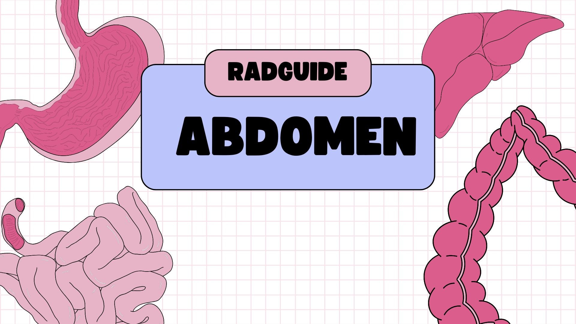
Radguide
The Abdomen : Osteology & Radiographic Technique
The Abdomen : Osteology & Radiographic Technique
Couldn't load pickup availability
Master Abdominal Anatomy, Osteology & Radiographic Technique with Confidence
Build a foundational understanding of the abdominal region with RadGuide’s comprehensive digital presentation. Designed for anatomy students, radiography trainees, and healthcare professionals, this resource offers a structured introduction to the skeletal framework, internal organ anatomy, and radiographic techniques essential for accurate abdominal imaging.
📘 In this presentation, you’ll explore:
Abdominal Osteology Overview
🦴 Skeletal Framework of the Abdominal Region
-
In-depth examination of the lumbar spine, lower ribs, pelvis, and sacrum as the bony structures that form the support for the abdominal cavity
-
Emphasis on anatomical landmarks such as the iliac crests, ASIS, and sacroiliac joints, which are crucial for proper patient positioning during abdominal imaging
-
Spatial relationships between the bones and soft tissues, aiding in understanding radiographic projections
Internal Abdominal Anatomy
🫀 Organs of the Abdominal Cavity
-
Overview of key internal organs, including the liver, stomach, intestines, spleen, kidneys, and pancreas, with details on their relative positions within the abdominal cavity
-
Understanding the anatomical relationship between organs and skeletal structures to optimize imaging positioning
-
How to recognize organs' contours and shadows in radiographs
Anatomical Regions and Quadrants
📏 Dividing the Abdomen for Clinical Relevance
-
Introduction to the abdominal regions and quadrants (e.g., RUQ, LUQ, RLQ, LLQ), and how these divisions assist in clinical examination and diagnosis
-
The importance of these regions in radiographic techniques, allowing for precise localization of pathologies like gallstones, appendicitis, or renal stones
-
Clear explanations of how these anatomical divisions correspond with radiographic projections to improve diagnostic clarity
Radiographic Techniques for Abdominal Imaging
📸 Standard Radiographic Views for the Abdomen
-
Step-by-step guidance on abdominal imaging techniques using standard projections such as AP supine, AP erect, and lateral decubitus
-
Key positioning tips for obtaining optimal abdominal radiographs, including proper center point placement, beam angle, and patient orientation
-
Radiographic considerations for imaging the abdomen in trauma cases, bowel obstruction, and fluid levels
🎯 Optimizing Radiographic Technique for Abdominal Imaging
-
Techniques for adjusting exposure factors (kVp, mAs) to ensure optimal soft tissue detail and bone structure visibility
-
Best practices for positioning to minimize overlapping shadows and ensure accurate visualization of internal organs and skeletal structures
-
Imaging techniques to highlight specific conditions, such as gastrointestinal tract issues, kidney stones, and abdominal trauma
💡 Clinical Imaging Considerations
-
How to assess abdominal pathologies like perforations, inflammatory conditions, and neoplasms using abdominal radiographs
-
Radiographic evaluation of the kidneys, liver, and spleen for conditions such as enlargement, calcifications, and masses
-
Advanced techniques for contrast-enhanced imaging in abdominal studies (e.g., IVP, CT scans) and their clinical relevance
Structural Relationships and Clinical Relevance
🔬 Anatomical Interactions and Pathological Relevance
-
Understanding how bones and soft tissues work together to protect internal organs, support posture, and interact in clinical assessments
-
Key focus on how to interpret radiographic findings in relation to common abdominal pathologies like hernia, gallstones, and pancreatitis
-
Clinical relevance of abdominal x-rays in emergency settings, such as acute abdomen, obstruction, and trauma cases
Educational Value and Application
Designed for:
-
Anatomy students and radiography trainees looking to solidify their understanding of abdominal anatomy and radiographic technique
-
Healthcare professionals involved in abdominal imaging or requiring an enhanced understanding of abdominal pathologies
-
Radiography educators teaching abdominal anatomy and imaging principles in clinical settings
Format: Digital Presentation (PowerPoint/PDF)
Delivery: Sent via email upon purchase
Level: Beginner to intermediate
Created by: Registered radiographer with clinical teaching experience
Share
