Radguide
Shoulder & Humerus : Radiographic Technique
Shoulder & Humerus : Radiographic Technique
Couldn't load pickup availability
Master Shoulder and Humerus Radiographic Techniques
This comprehensive PowerPoint presentation is designed to help radiography students, educators, and early-career radiographers gain proficiency in imaging the shoulder and humerus. The presentation covers essential radiographic techniques, from proper positioning to image evaluation, ensuring high-quality diagnostic images for accurate assessment.
📸 What’s Inside:
🦴 Anatomy of the Shoulder and Humerus
-
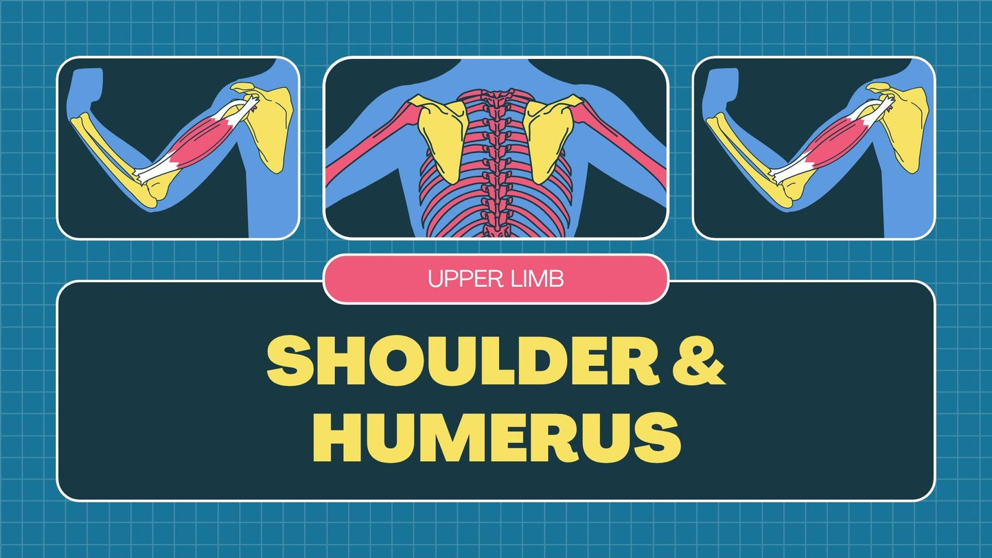
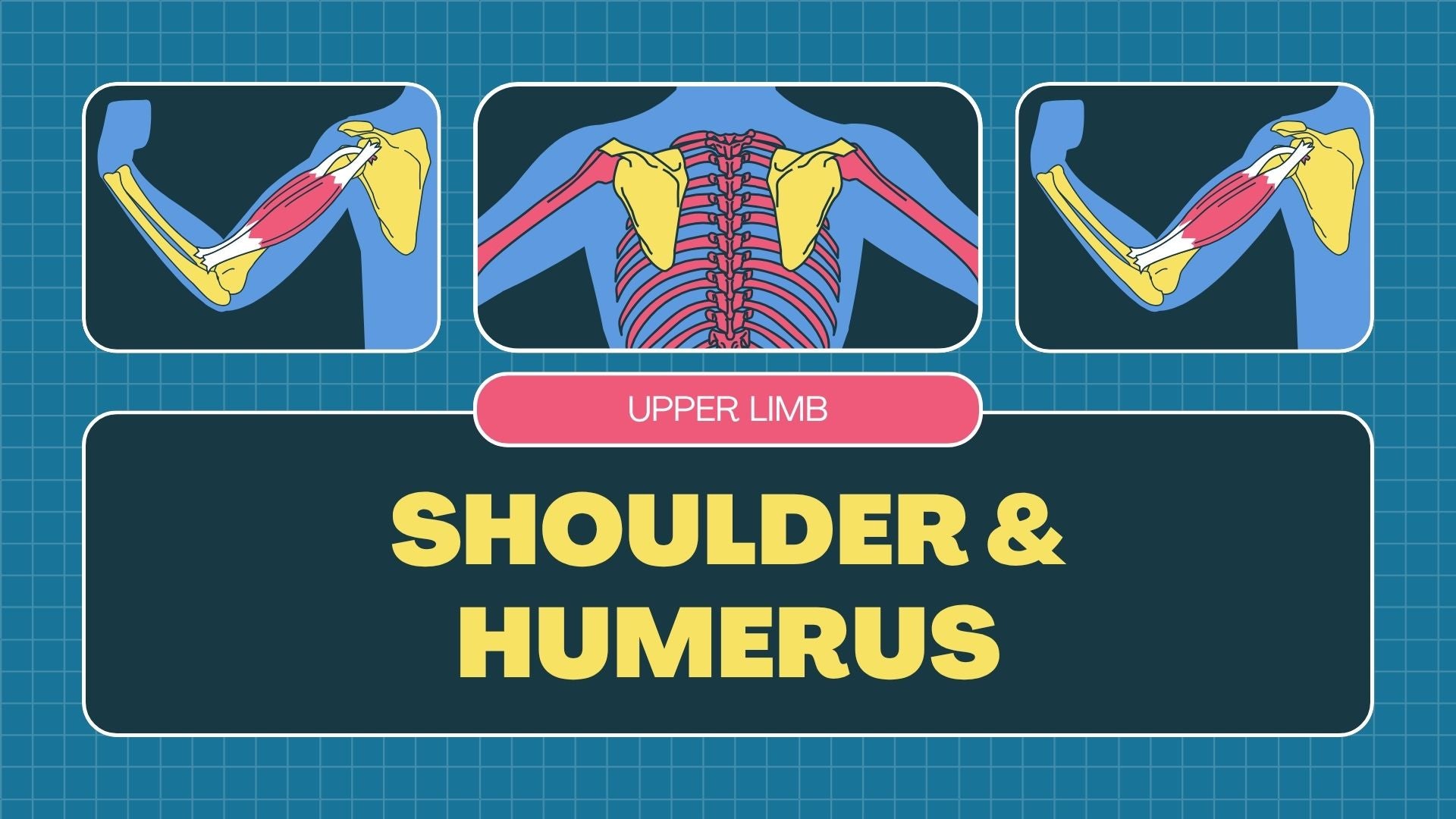
Overview of key shoulder structures: clavicle, scapula (including the acromion and glenoid), and humeral head
-
Detailed examination of the humerus, including its shaft, proximal features, and anatomical landmarks
-
Visual aids highlighting relevant bony structures for precise positioning
📸 Radiographic Techniques for the Shoulder and Humerus
-
Standard projections: AP shoulder, lateral (internal and external rotation), scapular Y view, and humerus views (AP, lateral)
-
Step-by-step guidance on patient positioning, centring points, and beam angulation
-
Tips for achieving clear, diagnostic images and reducing common positioning errors
🎯 Optimizing Radiographic Quality
-
Practical tips for selecting exposure factors and ensuring correct positioning
-
How to assess image quality and identify positioning flaws for improved diagnostic outcomes
-
Clinical indications for each projection and how to adapt for specific cases
💡 Ideal For:
-
Radiography students studying upper limb imaging techniques
-
Educators teaching shoulder and humerus radiography
-
Radiographers looking to refine their imaging skills and technique
Format: Digital presentation (PowerPoint/PDF)
Delivery: Sent via email upon purchase
Level: Beginner to intermediate
Created by: Registered radiographer with clinical teaching experience
Share


